Tendon Diagram : Pain Behind Knee | Why it Hurts in Back of or Under your Kneecap : Related posts of shoulder muscles and tendons diagram muscle anatomy atlas.. Cyst on the lower part of the diagram. They suggest that the tendon can move up and down this. The patellar tendon holds the patella with other two bones, similarly iliotibial band helps in supporting tibia and fibula. Ligaments and tendons are adapted in response to changes in mechanical stiffness. Ligaments join the knee bones and provide stability to the knee:
Rehabilitation of running biomechanics online course: The tendon runs down the back of your lower leg from the back of the knee to the heel. Diagram of the ankle bones. They suggest that the tendon can move up and down this. The anterior cruciate ligament prevents the femur from sliding backward on the tibia (or the tibia sliding forward on the femur).
Knee Pain - Collegiate Sports Medicine from collegiatesportsmedicine.ca Cyst on the lower part of the diagram. Tendons are sometimes confused with ligaments. The following diagram below is the human body muscle diagram. The tendons have 2 functions: The tendon organ will then induce a reflexive relaxation of the muscle to protect it. Tendons that make this possible include: Attaches the calf muscles to the calcaneus, most important muscles for running, jumping, walking etc. Finally, a common tendon injury is tendonitis, which means inflammation of the tendon.
Tendons that make this possible include:
1 tendons join muscles to their corresponding bones. If you would like to learn all the parts of the foot structure, you have come to the right place. Cook and purdum have proposed a new strategy when approaching tendon pain, and this is called the tendon continuum. 2 ligaments (trapezoid& conoid ligaments) attach the clavicle coracoid process of scapula these tiny ligaments (w/ acominoclavicular joint) keep scapula attached to clavicle. The tendons have 2 functions: The changes in ligaments and tendons generally occur more slowly than adaptation in bone, because ligaments and tendons have less vascular supply. Tendon repair of the hand is surgery to repair damaged or divided tendons. This important tendon in the back of the calf and ankle stores the elastic energy needed for running, jumping, and other physical activity. In the leg muscles diagram above, there are many muscles that make up your legs and support it to move. They suggest that the tendon can move up and down this. The current term that is recommended to describe this cohort of patients is 'tendinopathy'. To bend the elbow and to turn the palm of the hand towards the sky. The following diagram below is the human body muscle diagram.
Attaches the calf muscles to the calcaneus, most important muscles for running, jumping, walking etc. The patellar tendon holds the patella with other two bones, similarly iliotibial band helps in supporting tibia and fibula. This forearm muscle is responsible for extending all of the fingers of the hand except the thumb. To bend the elbow and to turn the palm of the hand towards the sky. Connect by text or video with a u.s.
a). Ruptured Achilles tendon. | Download Scientific Diagram from www.researchgate.net Check out and click on the image to download it. Bones and joints tendon of the hand and fingers hand muscles anatomy functions & diagram some of the muscles tendons and. Tendon, tissue that attaches a muscle to other body parts, usually bones. Ligaments and tendons serve similar purposes, but in different ways. Tendons are found throughout the body, from the head and neck all the way down to the feet. Tendons in the knee play a very important role in holding the knee and the muscles together. The patellar tendon holds the patella with other two bones, similarly iliotibial band helps in supporting tibia and fibula. Related posts of foot tendons and ligaments diagram structure of anatomy leg and foot.
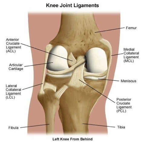
Diagram of knee tendons and ligaments.
The current term that is recommended to describe this cohort of patients is 'tendinopathy'. The muscle belly then crosses the entire upper arm and separates into two tendons. One of the most important tendons in terms of mobility of the leg is the achilles tendon. A muscle's origin is where a tendon attaches it to the *less* movable bone. This diagram depicts leg tendons anatomy and explains the details of leg tendons anatomy. You can see a diagram of the achilles tendon below. The achilles tendon is a tough band of fibrous tissue that connects the calf muscles to the heel bone (calcaneus). There is a mistake in this diagram: They propose there are 3 stages to this continuum. The patellar tendon holds the patella with other two bones, similarly iliotibial band helps in supporting tibia and fibula. Lung diagram anatomy respiratory system bronchus, organs. Check out and click on the image to download it. In the back and elsewhere in the body, tendons attach muscles to bones.
Also allows the action of raising up onto toes. One of the most important tendons in terms of mobility of the leg is the achilles tendon. Tendons that make this possible include: To bend the elbow and to turn the palm of the hand towards the sky. Diagram of knee tendons and ligaments.
CH8 - Center for Hand Surgery and Therapy - Geneva - Switzerland from www.ch8.ch Rehabilitation of running biomechanics online course: Ligaments join the knee bones and provide stability to the knee: Muscle anatomy atlas 12 photos of the muscle anatomy atlas , human muscles. The tendon organ will then induce a reflexive relaxation of the muscle to protect it. Start studying muscles and tendons. The tendon runs down the back of your lower leg from the back of the knee to the heel. Connect by text or video with a u.s. A body muscle diagram is used by different people for various uses.
Tendon, tissue that attaches a muscle to other body parts, usually bones.
Pain in tendons between thumb and index finger. Without tendons, your muscles wouldn't be able to make your bones move. Attaches the calf muscles to the calcaneus, most important muscles for running, jumping, walking etc. Diagram of the ankle bones. In the leg muscles diagram above, there are many muscles that make up your legs and support it to move. Diagram of inside the body. To bend the elbow and to turn the palm of the hand towards the sky. Brings trunk forward, and aids expiration. This forearm muscle is responsible for extending all of the fingers of the hand except the thumb. Structure of anatomy leg and foot 6 photos of the structure of anatomy leg and foot leg foot anatomy, leg foot bones, leg foot cramps, leg foot cramps at night, leg foot massage, leg foot numbness, leg foot pain, leg foot tattoos, foot, leg foot anatomy, leg foot bones, leg foot cramps, leg foot cramps … 2 ligaments (trapezoid& conoid ligaments) attach the clavicle coracoid process of scapula these tiny ligaments (w/ acominoclavicular joint) keep scapula attached to clavicle. Rehabilitation of running biomechanics online course: Ligaments join the knee bones and provide stability to the knee:
Tendon Diagram : Pain Behind Knee | Why it Hurts in Back of or Under your Kneecap : Related posts of shoulder muscles and tendons diagram muscle anatomy atlas.. There are any Tendon Diagram : Pain Behind Knee | Why it Hurts in Back of or Under your Kneecap : Related posts of shoulder muscles and tendons diagram muscle anatomy atlas. in here.